
Most of what failed me in the first decade of hEDS and MCAS care was sequencing. The right therapy in the wrong order is still the wrong therapy. This is the order that finally worked.
Hypermobile Ehlers-Danlos syndrome (hEDS) and mast cell activation syndrome (MCAS) are multisystem disorders that overlap so often that treating one without the other usually means treating neither well. hEDS produces connective tissue fragility, joint hypermobility, skin hyperextensibility, and a constant low rate of tissue injury. MCAS adds mast cells releasing excessive inflammatory mediators — histamine, tryptase, matrix metalloproteinase-9 (MMP9) — on top of that fragile substrate. Layer in dysautonomia (POTS), fibromyalgia, and the chronic inflammation often called inflammaging, and you have a system that degrades itself faster than it can repair.
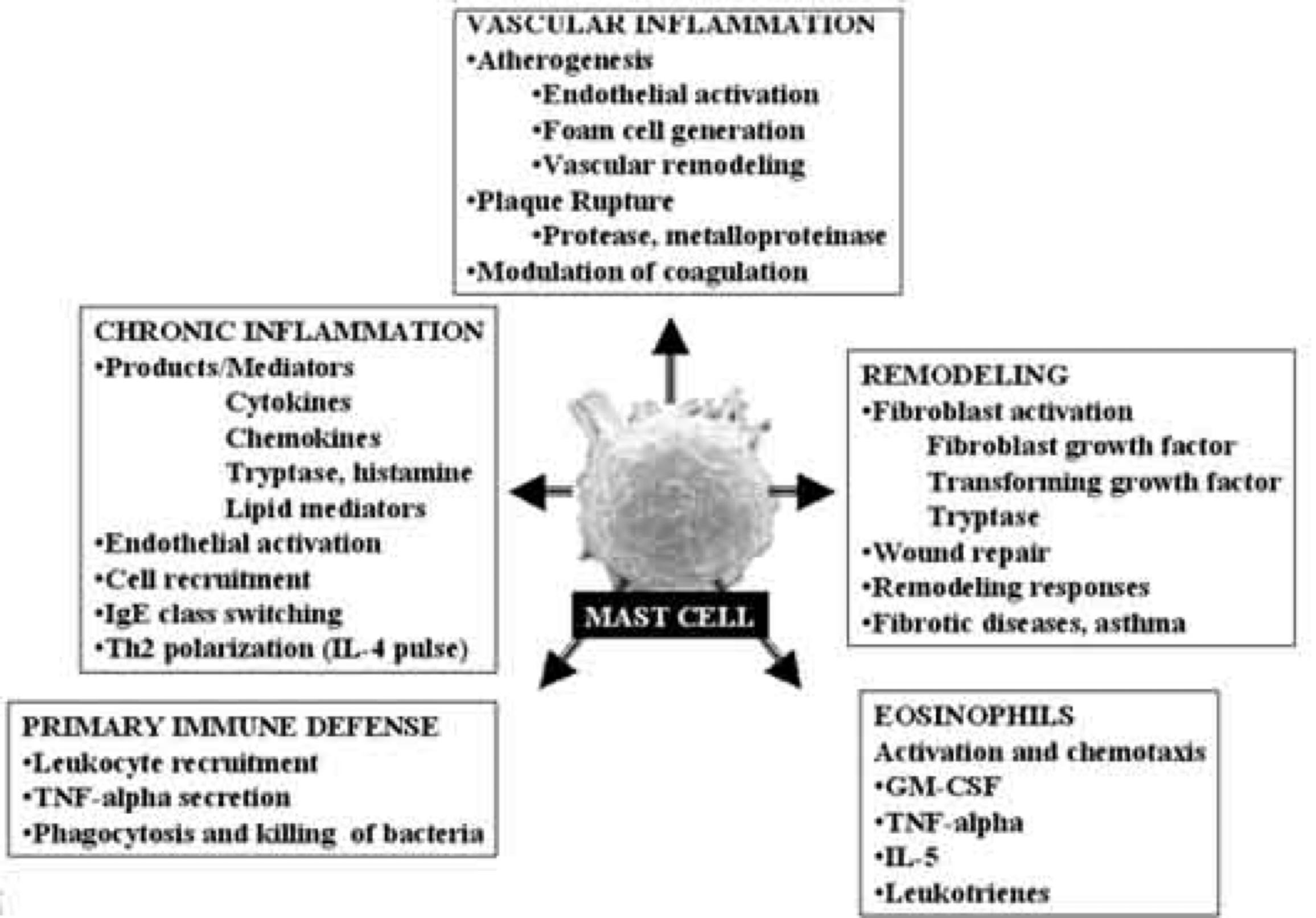
This is the protocol I run, in the order I run it.
1. Diagnostics before anything else
Clinical and laboratory workup
Document everything: chronic pain, hypermobility, fatigue, GI involvement, dysautonomia (POTS), allergic and anaphylactoid events. The history is the foundation; nothing downstream works without it.
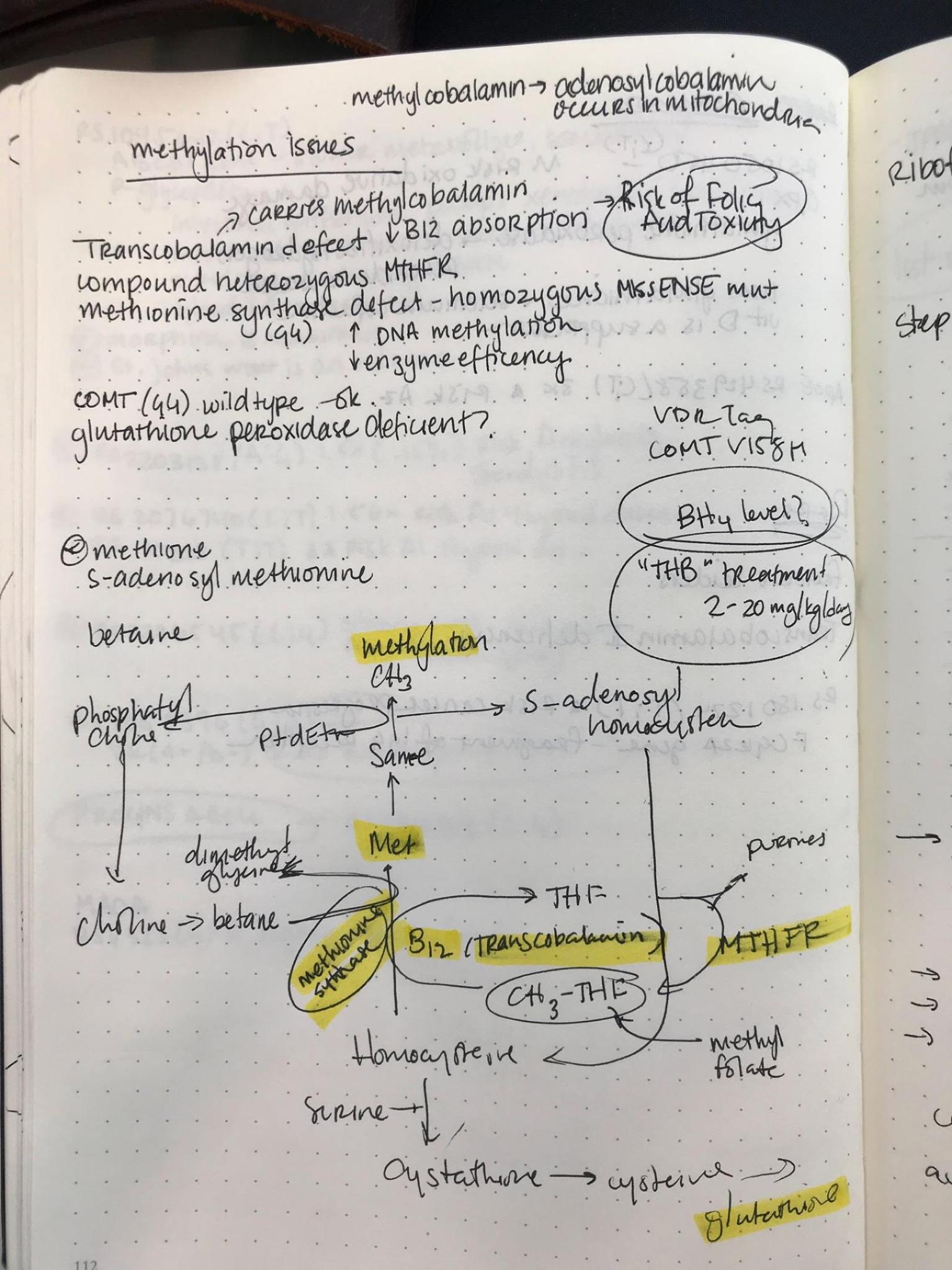
Genetic testing. There is no single genetic marker for hEDS, but collagen and connective-tissue mutations are commonly found and clinically informative. My own panel returned COL5A2: c.1633C>T, COL4A4: c.3979G>A, and COL17A1: c.4304C>T (p.A1435V) (Afrin et al., 2016). Individually many of these variants are labeled benign. Stacked, they are not.
MCAS biomarkers. Serum tryptase, histamine, prostaglandin D2, and especially MMP9. Elevated MMP9 during flares is the cleanest objective signal of active collagen breakdown I have found (Theoharides et al., 2020).
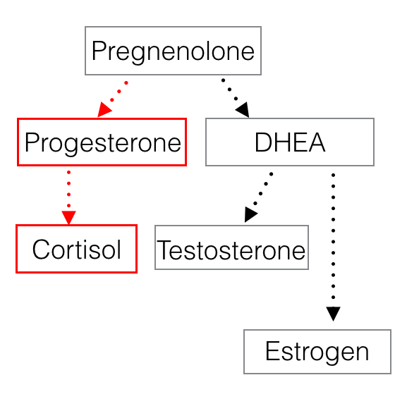
Endocrine and metabolic panels. Chronic mast-cell driven stress drives high cortisol, high sex hormone-binding globulin (SHBG), and low free testosterone. The result is impaired recovery, fatigue, and a body that can’t keep up with its own damage. Measure total and free testosterone, SHBG, cortisol, and pregnenolone. The “pregnenolone steal” — where cortisol production cannibalizes anabolic precursors — is real and addressable.
- If pregnenolone is low, supplement pregnenolone and boron (boron binds SHBG).
- Pregnenolone is the precursor to aldosterone. Nocturia (frequent nighttime urination) is the practical clinical marker. Titrate up in 30 mg increments to 90 mg until nocturia resolves and full-night sleep returns. Always pair with boron so SHBG doesn’t spike further. Taper, never stop suddenly. Increase the dose during high-stress periods, injury, or sleep loss.
Autonomic testing. If a patient describes arrhythmia but a standard EKG is clean, get a Holter monitor.
What clinicians should take away
Connective tissue and methylation mutations don’t just support a hEDS diagnosis — they can exacerbate MCAS by impairing baseline tissue repair (Molderings et al., 2017). MMP9 is the most useful single biomarker I monitor for flare intensity and ongoing collagen degradation.
2. Injury management and rehabilitation, in order
A. Stabilize the neck and SI joints first
The neck and SI joints are upstream of almost everything else. Stabilize them first or you will chase symptoms downstream for years.
Neck. Image with flexion-extension and rotational MRI — atlantoaxial instability and cervical compromise do not show up reliably on static scans. Then get to a regenerative spine specialist early. Dr. Marko Bodor is the expert I trust for facet joints, though he is not taking on complex CCI cases at the moment and I am working on updated referrals.
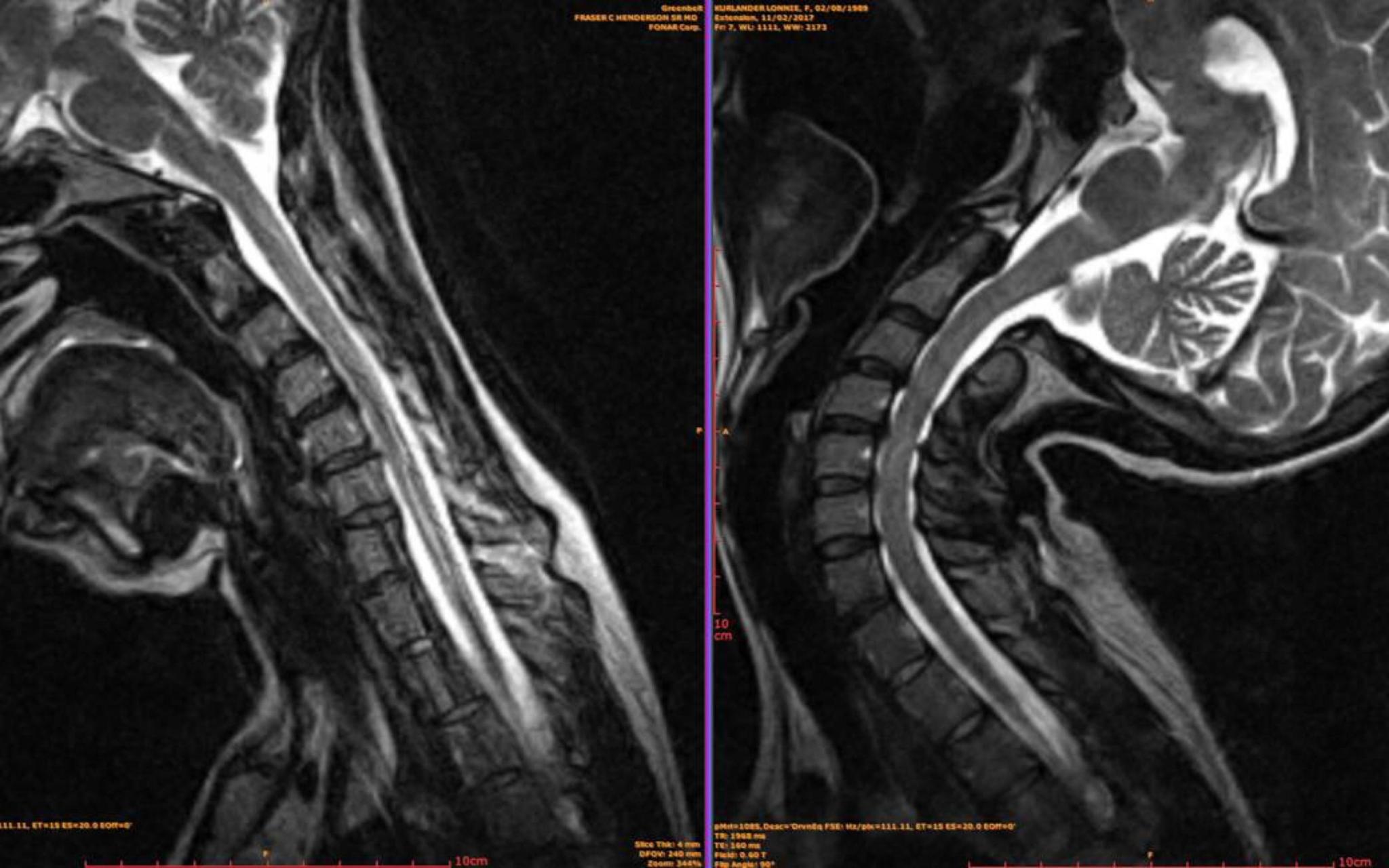
A few things I want patients and providers to understand about cervical sequencing:
-
Treating posterior ligaments without addressing the anterior ligaments can create imbalance. A holistic approach matters for good candidates.
-
Chris Centeno reports that in his PICL randomized controlled trial, 70% of participants did not proceed to surgical intervention. I do not know the exact time frame for that figure, and the inclusion criteria are quite strict, so generalization to broader populations is uncertain. There is also active debate about whether anterior pannus or compression is a contraindication for PICL; I walk through that question in detail here.
-
Vertebral slippage visible on static (non-motion) imaging may favor surgical stabilization over regenerative approaches.
-
Centeno is the only provider I know of with deep experience treating the anterior ligaments. Bodor and Centeno have cross-trained — Bodor on facets, Centeno on anterior. A user-maintained list of other alar-ligament injectors exists on Reddit; I have not vetted it. A good doctor knows what they do not know.
-
If you need surgery, my own was with Dr. Fraser Henderson almost a decade ago, my outcome is good, and we are back in touch. I have an additional unverified recommendation for Dr. Neill Wright that I need to investigate further.
-
Anyone considering surgery or regenerative work should first be evaluated by an expert DO for spinal curve and SI joint placement. A lost curve or displaced SI joint will tug on the spinal cord and undermine everything downstream.
-
Even if you proceed with surgery, stabilize it with regenerative medicine to prevent failure. Rehab is a lifestyle, not a phase.
SI joints. SI joint dysfunction is endemic in hEDS. Initial treatment is regenerative stabilization plus mobilization with a skilled, gentle osteopathic physician. PRP, PRF, PRGF, BMAC, and other orthobiologics — and where regulation permits, stem cells and exosomes — can be used alone or in combination. Cells and exosomes are not FDA-approved, but the literature is real and worth understanding:
- Clinical use of stem cells in orthopaedics — European Cells and Materials
- Clinical applications of stem cell-derived exosomes — Nature
- Exosomes in clinical trials — Cell Communication and Signaling
The Cranial Academy is a good place to find a competent local DO. Practitioners I rely on or recommend: Dr. Dan Shadoan, Dr. Matthew Gilmartin, Dr. Vivian Levy, Dr. Edward Edris, Dr. Nick Andrews, and Dr. Scott Corbett. I plan to organize regional endorsements over time.
B. Then move outward
Once the neck and SI joints are stable, work outward to the big joints (hips, knees, shoulders), then to hands and feet. Before assuming “tethered cord,” confirm SI joint position — a displaced SI joint pulls on the lower spinal cord and mimics tethered cord symptoms.
Find a regenerative practitioner you can see regularly. Dr. Rowan Paul is one example — his practice is over 80% EDS, including elite athletes, Olympians, and professional ballerinas.
C. Rehab and strength
- Begin with low-impact work. It may take 3-5 years to safely reach heavy weight training. That is fine.
- Impact sports (running) are typically not tolerated. Progress slowly to heavy weights without pain. Never push through popping, clicking, or pain — back off, see your DO and regenerative specialist, and reset.
- Avoid excessive axial loading if you have disc issues, slippage, or implants (artificial discs).
- Emphasize functional movement, flexibility, and proprioception.
- Bodyweight → resistance → loaded, always monitoring joint stability.
- Setbacks and flares are inevitable. Have a plan for them.
D. Adjunctive interventions that earn their place
Stellate ganglion blocks. These produce some of the most durable improvements I have seen in dysautonomia and in the longevity of regenerative procedures. The most complete protocol I have found is the Complete Neuro Reset with Dr. Jonathan Kuo in New York. Dr. Matthew Cook in the Bay Area is the closest competitor. Both protocols integrate vagal and pharmacologic layers beyond the block itself.
Hyperbaric oxygen. Synergizes with regenerative treatments to accelerate repair, particularly useful for serious neck injuries and large tears, and it reduces systemic inflammation. It is not without risk — cataracts are a known consequence of overuse.
Peptides and orthobiologics. PRP, exosome therapy, and peptide therapies (TB-500, thymosin alpha, BPC-157) for tissue repair and inflammation. The Peptide Sciences gut formula has been a sustained help for me. Address dysbiosis alongside this with a functional medicine physician.
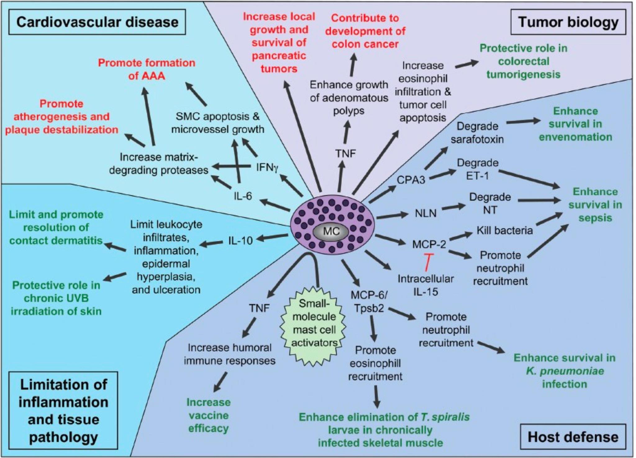
3. Controlling mast cell activity
Acute flares (IV protocol)
- Diphenhydramine (Benadryl) — H1 blocker, fast histamine control. Use with awareness of cognitive side effects over time; aim to reduce dependence as the system stabilizes. Exosomes may help mitigate long-term cognitive cost; I am watching that literature.
- Famotidine (Pepcid) — H2 blocker, complements H1 blockade and reduces gastric acid.
- Lysine and proline — collagen substrate.
- Glutathione and vitamin C — antioxidants. Vitamin C is also a potent mast cell stabilizer.
- Toradol (ketorolac) — only if tolerated, and never immediately after regenerative procedures. NSAIDs impair tissue repair.
Chronic oral and topical
- H1/H2 blockade. Start with a non-sedating H1 plus an H2. Run a combination for 2-3 weeks; if benefit isn’t clear, rotate one component. Iterate until you find a pair that produces a noticeable shift in baseline symptoms.
- Ketotifen. A mast cell stabilizer with good efficacy in many patients.
- Celecoxib. COX-2 selective, lower GI risk — but still an NSAID and should not be combined with active regenerative treatment.
- Sertraline. Stabilized my symptoms substantially. Several SSRIs modulate mast cell behavior.
- Sodium cromolyn. Not universal, but works in a subset.
The longer list — Dr. Lawrence Afrin is among the foremost MCAS experts and now works inside a functional medicine practice. Dr. Stephanie Daniel at Functional Medicine SF is another strong functional medicine option. Functional medicine is central to building a working medication and trigger-elimination plan.
- Low-dose imatinib has been effective in some patients.
- Continuous IV diphenhydramine infusion has helped severely affected patients in near-constant anaphylaxis. It is inpatient and calibrated in hospital. If you need this, you need Afrin — he is the only physician I know of with that depth of experience.
Lifestyle and trigger management
- Environmental control. Identify and avoid triggers — mold (double-bag and properly dispose of contaminated items), food additives, cold, stress.
- Allergy testing. Evaluate IgG and IgA sensitivities. Carry trigger-avoidance cards to prevent cross-contamination.
- Diet and supplements.
- Thorne Methylgard Plus (or equivalent) if you have methylation mutations.
- Pregnenolone 30-90 mg with boron when cortisol is high (Agarwal et al., 2020).
- Lysine for collagen support.
- Protein and caloric adequacy. High-quality protein supports muscle, tissue repair, and temperature regulation.
- Quercetin and fish oil for mast cell and inflammatory modulation.
- Nattokinase for vascular support.
Monitoring
- MMP9 during flares — objective signal of collagen breakdown (Kohler et al., 2019).
- Free testosterone, SHBG, cortisol regularly to catch hormonal drift driving flares.
4. The integration: why sequencing matters
Genetically weakened connective tissue plus episodic mast cell-driven inflammation produces a double hit on tissue integrity. MMP9 actively degrades collagen; histamine drives vasodilation and increased venous compliance, worsening dysautonomia and POTS (Theoharides et al., 2020; Raj et al., 2019).
Therefore:
- Stabilize the neck and SI joints before larger joints and extremities. If atlantoaxial or craniocervical instability is suspected — or a functional Chiari appears on motion imaging — get into a properly fitted cervical collar immediately and address the neck first. My arrhythmias resolved when I addressed my neck.
- Don’t combine NSAIDs (Toradol, celecoxib) with active regenerative treatment.
- Always screen for metabolic and hormonal imbalance in EDS/MCAS patients.
- Stellate ganglion blocks and hyperbaric oxygen are real adjuncts, not luxuries (Weinstock et al., 2021).
A working roadmap
- Diagnose thoroughly. Genetic panel, mast cell mediators including MMP9, endocrine panel, allergy testing, detailed symptom log.
- Stabilize the spine. Neck and SI joints first, with regenerative treatment, injections, or surgery if necessary. Find a practitioner you can see regularly.
- Rebuild systematically. Controlled non-axial work, then bodyweight, then weighted. Protein-forward diet. Use stellate blocks and HBOT where indicated.
- Control mast cells. H1/H2, ketotifen, selective anti-inflammatories. IV protocol during flares. Avoid NSAIDs immediately around regenerative work. Watch MMP9. Prioritize sleep. Keep dye-free oral Benadryl on you. Make a business card with your allergies and cross-contamination protocols on the back — restaurant staff appreciate it and you reduce error.
- Optimize endocrine and metabolic state. Free testosterone, SHBG, cortisol, pregnenolone. Supplement as indicated (pregnenolone, boron, Thorne Methylgard Plus, SAM-e, lysine).
- Adjust your environment. Eliminate triggers (food additives, mold, temperature extremes, stress). Detox protocols (activated charcoal, chlorella) when indicated. Treat rehab as a daily lifestyle.
- Iterate. Document with regular follow-up and biomarker tracking. Adjust exercise based on current strength, stability, and pain. Build an interdisciplinary team — rheumatology, immunology, regenerative medicine, PT — and refine continuously.
Closing
Managing hEDS and MCAS is not a single intervention. It is a coordinated, ordered, evidence-driven approach that respects both genetic vulnerability and inflammatory dynamics. My own path — from debilitating instability and multisystem pain to ten years of progressive restoration of strength and function — is not a miracle and not luck. It is the result of sequencing, biomarkers, and a team that takes the underlying biology seriously.
Further reading
References
- Afrin LB, et al. “Often seen, rarely recognized: mast cell activation disease — a guide to diagnosis and therapeutic options.” Ann Med. 2016;48(3):190-201. DOI: 10.3109/07853890.2016.1161231
- Molderings GJ, et al. “Risk of solid cancer in patients with mast cell activation syndrome: results from Germany and USA.” F1000Res. 2017;6:1889. DOI: 10.5256/f1000research.12730.1
- Karsenty G, Wagner EF. “An integrated model of skeletal development and bone homeostasis.” Science. 2002;296(5565):1821-1824.
- Theoharides TC, et al. “Mast cell activation syndrome: diagnostic criteria, and a practical approach to diagnosis and management.” J Allergy Clin Immunol Pract. 2020.
- Franceschi C, et al. “Inflammaging: a new immune-metabolic viewpoint for age-related diseases.” Nat Rev Endocrinol. 2018;14(10):576-590. DOI: 10.1038/nrendo.2018.56
- Raj SR, et al. “Pathophysiology of postural orthostatic tachycardia syndrome.” Circulation. 2019. DOI: 10.1161/CIRCULATIONAHA.118.038493
- Kritas SK, et al. “Mast cells in COVID-19: the silent players.” J Allergy Clin Immunol. 2020.
Not medical, legal, or regulatory advice. As of May 2026: no exosome product has FDA approval for therapeutic use; several cell therapies are FDA-approved (HSCT, CAR-T, sickle-cell gene editing, Ryoncil for graft-versus-host disease) but none for the orthopedic or regenerative-medicine indications discussed here. A few states — Texas, Florida (effective July 2025), Utah, Mississippi — have created limited permission frameworks for certain non-FDA-approved stem cell therapies; none extend to exosomes, and federal law still preempts. Full regulatory rundown →